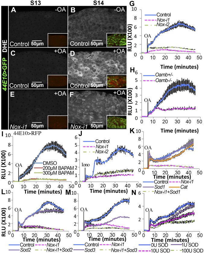
Fig. 3.
OA activates NOX to produce superoxide extracellularly. (A–F) Representative images show DHE staining (white in A–F) in control (A–D) and Nox-i1 (E and F) follicles after 30-min culture without (A and B) or with (C–F) OA stimulation. The Insets are low-magnification images with 44E10>GFP expression (green, marking stage-14 follicles) and DHE staining (red). (G–N) l-012 Luminescence-dependent O2•− quantification in mature follicles stimulated with OA (G–I and K–N) or ionomycin (J) at the 5-min time point. Mature follicles with different genotypes were isolated according to 44E10>RFP expression. Mature follicles in I were pretreated with BAPTA-AM for 30 min before l-012 detection. Mature follicles in N were supplemented with SOD extract from bovine erythrocytes in the culture medium. Iono, ionomycin; Nox-i, Nox-RNAi; RLU, relative luminometer unit.

