Fig. 1.
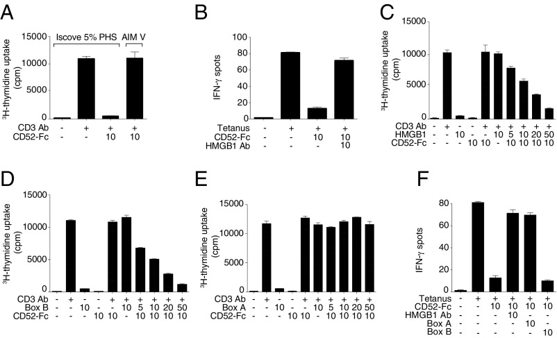
T cell suppression by soluble CD52 requires HMGB1 Box B and is antagonized by HMGB1 Box A. (A) Proliferation of human PBMCs incubated in serum-containing IP5 medium (Iscove’s modified Dulbecco’s medium with 5% PHS) or serum-free medium (AIM V) for 48 h alone or with plate-bound anti-CD3 antibody (1 μg/mL) and CD52-Fc. (B) IFN-γ production measured by ELISpot from human PBMCs (1 × 10⁵) incubated in serum-containing medium with no antigen or tetanus toxoid (10 Lfu/mL) in the presence of CD52-Fc and neutralizing antibody to HMGB1. (C) Proliferation of human PBMCs incubated in serum-free medium alone or with plate-bound anti-CD3 antibody (1 μg/mL), CD52-Fc and with increasing concentrations of disulfide-HMGB1, or HMGB1 Box B (D), or HMGB1 Box A (E). Proliferation was measured as [3H] thymidine uptake. (F) IFN-γ production measured by ELISpot from human PBMCs (1 × 10⁵) incubated in serum-containing medium in the presence of HMGB1 Box A or HMGB1 Box B recombinant proteins and neutralizing antibody to HMGB1. Concentrations of recombinant proteins in every figure are in μg/mL. Serum-free medium was used in experiments in A, C, D, and E.

