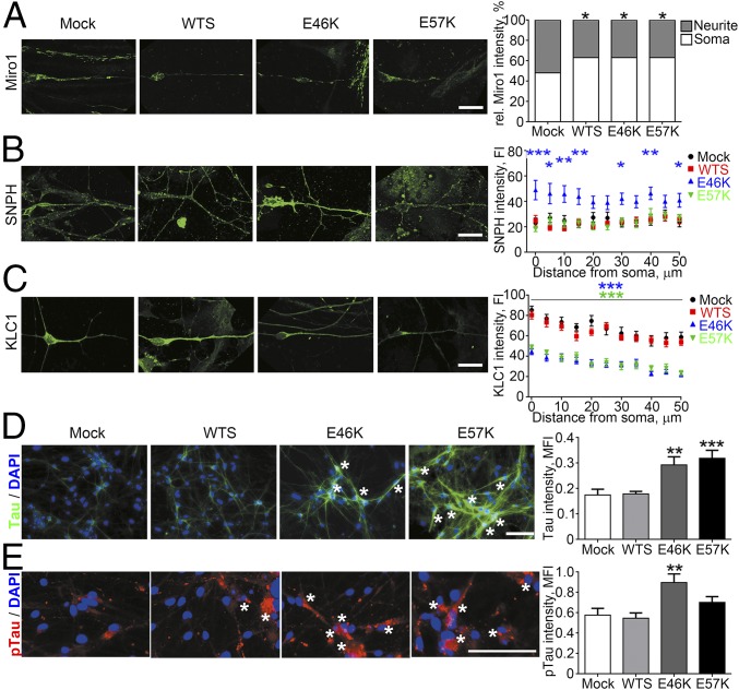
Fig. 3.
Spatial distribution and expression of kinesin adaptor proteins is profoundly altered in the presence of α-Syn oligomers. (A) Significantly decreased neurite-to-soma ratios of Miro1 were measured in WTS, E46K, and E57K neurons. (B) A significant increase of SNPH was found in E46K neurites. (C) Significantly decreased levels of KLC1 were found in neurites of oligomer-containing E46K and E57K neurons. (D) Significantly increased total Tau levels (green) especially in a perinuclear region (asterisks) and (E) pTau (red) accumulations (asterisks) in E46K and E57K neurons compared with Mock and WTS. Tau and pTau levels were normalized to the number of DAPI-positive viable cells. Data are presented as mean ± SEM. MFI, mean fluorescence intensity. *P ≤ 0.05, **P ≤ 0.01, ***P ≤ 0.001. (Scale bars: 20 µm in A–C and 100 µm in D and E.)

