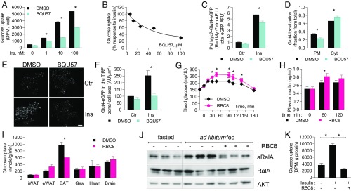
Fig. 1.
Ral inhibitors reduce glucose uptake and plasma membrane localization of Glut4. (A–G) 3T3-L1 adipocytes were pretreated with 100 μM BQU57 for 1 h and stimulated with 10 nM insulin for 30 min, unless indicated otherwise. (A and B) Glucose uptake in 3T3-L1 adipocytes treated with (A) 50 μM BQU57 and various concentrations of insulin and (B) dilutions of BQU57 and 10 nM insulin; four replicates for each condition. (C) Quantitation of Myc7-Glut4-EGFP localization in 3T3-L1 adipocytes; 22 cells per group. (D) Quantitation of endogenous Glut4 localization; 22 control and 25 BQU57-treated cells. (E and F) TIRF imaging of Myc7-Glyt4-EGFP in 3T3-L1 adipocytes 3 min after insulin administration (E) and its quantitation (F); five cells in each condition. (G–J) Ad libitum-fed lean mice were i.p. injected with 50 mg/kg RBC8. (G and H) Blood glucose (G) and insulin (H) levels; 11 mice per group. (I) 2-Deoxy-d-glucose-phosphate in tissues 40 min after RBC8 administration; eight mice per group. (J) WB of BAT lysates at 40-min time point. (K) 2-Deoxy-d-glucose uptake in primary brown adipocytes. The data are presented as mean ± SEM; *P < 0.05. aRalA, active RalA; Ctr, control; Cyt, cytosolic; PM, plasma membrane.

