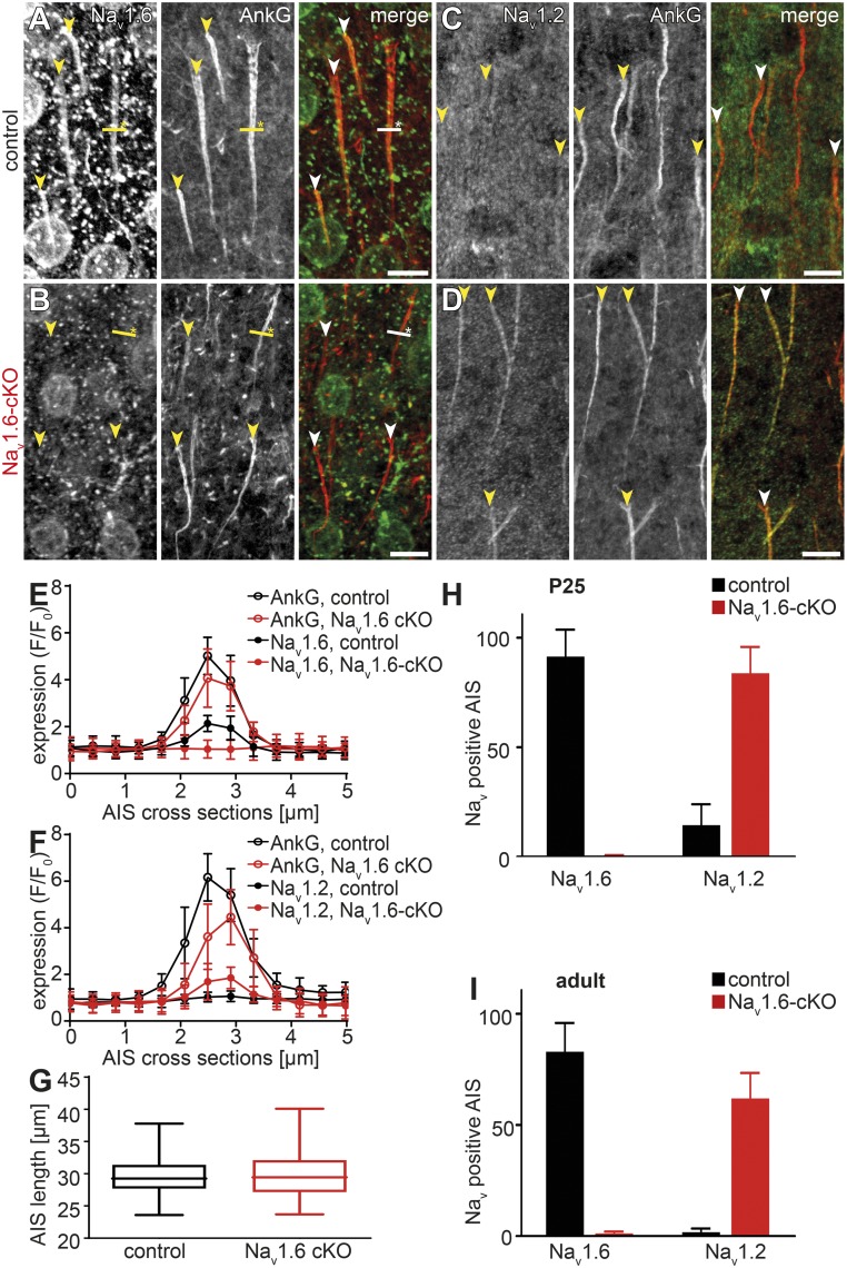
Fig. 1.
Expression of Na+ channel subtypes at the AIS of L5 pyramidal neurons in control and Nav1.6-cKO mice. (A–D) Confocal micrographs of Nav1.6 (A and B), Nav1.2 (C and D), and AnkG immunostains in the layer 5 cortical sections of adult control (A and C) and Nav1.6-cKO (B and D) mice. Arrowheads indicate the proximal boundary of the AnkG-labeled AIS. Yellow lines with asterisks indicate the cross-section positions analyzed in E and F. Nav1.6 channels are present over the entire AIS length in neurons of adult control mice, while they are absent in Nav1.6 cKO mice (A and B, merged images). Nav1.2 channels are barely detectable in AISs of control neurons, but strongly up-regulated in the Nav1.6-deficient neurons (C and D, merged images). (Scale bars, 10 µm.) (E and F) Sections across the AISs (2.5 µm on both sides) were used to quantify Nav1.6, Nav1.2, and AnkG expression in adult mice (analyzed as F/F0). While AnkG expression remained constant in control (black) and Nav1.6-deficient (red) AISs, Nav1.2 is present in the Nav1.6-cKO AISs at approximately the same levels as Nav1.6 in control AISs. (G) Analysis of AIS length as measured by AnkG expression did not reveal significant difference between Nav1.6-cKO (red) and control (black) neurons. (H and I) Fractions of Nav1.6- and Nav1.2-positive AISs of control (black) and Nav1.6-cKO (red) neurons in young (P25, H) and adult (P60, I) mice. Note that the Nav1.6 deficiency is almost completely compensated for by Nav1.2.

