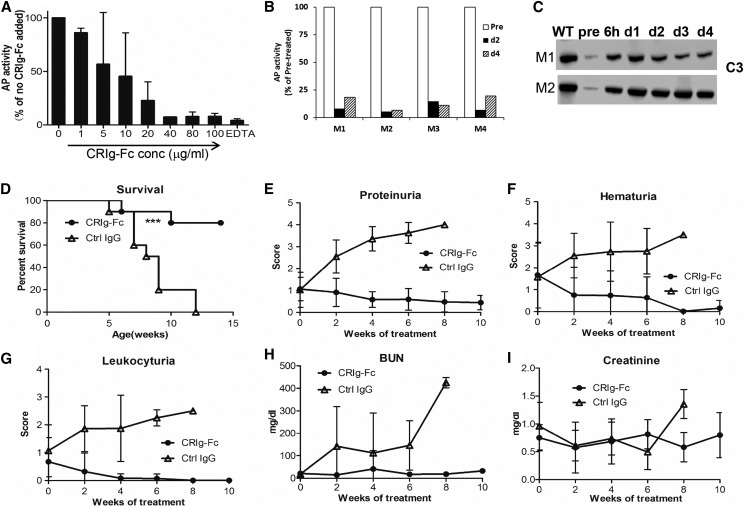
Figure 1.
CRIg-Fc inhibits AP activity in normal and mutant mice and prevents fatal C3G disease. (A) At 40 μg/ml or higher, CRIg-Fc effectively inhibited AP activity in 50% WT mouse serum. Data from two independent experiments (one with duplicate and the other triplicate assays) were pooled and plotted together. Results are presented as mean (SD). (B) CRIg-Fc inhibited AP activity in vivo in WT mice. Data from four WT mice (M01, M02, M03, and M04) were plotted individually. Mice were injected with 50 mg/kg CRIg-Fc and LPS-dependent AP activity was measured using 10% serum, in duplicate assays (average is shown). AP activity at day 2 and day 4 was normalized to that of the pretreatment sample (pre). (C) Western blotting shows that CRIg-Fc treatment inhibited C3 consumption and markedly elevated plasma C3 levels in two FHm/m mice (M1 and M2). Western blot band shown denotes intact C3 α-chain. A single dose of 1 mg CRIg-Fc was administered to each mouse. Blood samples were collected immediately before and at 6 hours and 1, 2, 3, and 4 days after CRIg-Fc injection. (D–I). Treatment of FHm/mP−/− mice with control IgG had no effect on survival (100% mortality) and C3G disease development as assessed by proteinuria, hematuria, leukocyturia, BUN, and serum creatinine. In contrast, treatment with CRIg-Fc significantly improved survival (80%) and multiple disease parameters. FHm/mP−/− mice received treatments (n=10 mice per group) starting at 4 weeks of age and lasting for 10 weeks (50 mg/kg, administered intraperitoneally). Data shown in (E–I) represent mean (SD). ***P<0.001, log-rank test. ctrl, control; conc, concentration.

