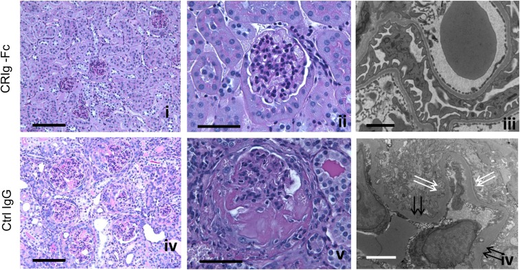
Figure 2.
FHm/mP−/− mice treated with CRIg-Fc but not isotype control IgG mAb had normal kidney histology. Period Periodic acid–Schiff staining shows that a representative control IgG–treated mouse developed fatal kidney damage with numerous crescents, significant glomerular enlargement, inflammation, and tubulointerstitial injury (D and E). In contrast, in a representative mouse treated with CRIg-Fc, only mild hypercellularity was observed in some glomeruli and no signs of significant crescents or tissue damage were evident (A and B). By electron microscopy, in a representative CRIg-Fc–treated mouse, normal GBM and intact podocyte foot processes (C) were seen. Signs of podocyte injury, irregular GBM thickening (F, double black arrows) and foot process effacement (F, double white arrows) were clearly present in a representative control IgG–treated FHm/mP−/− mouse. Bars, 100 μm in (A and D), 50 μm in (B and E), 2 μm in (C), and 4 μm in (F).

