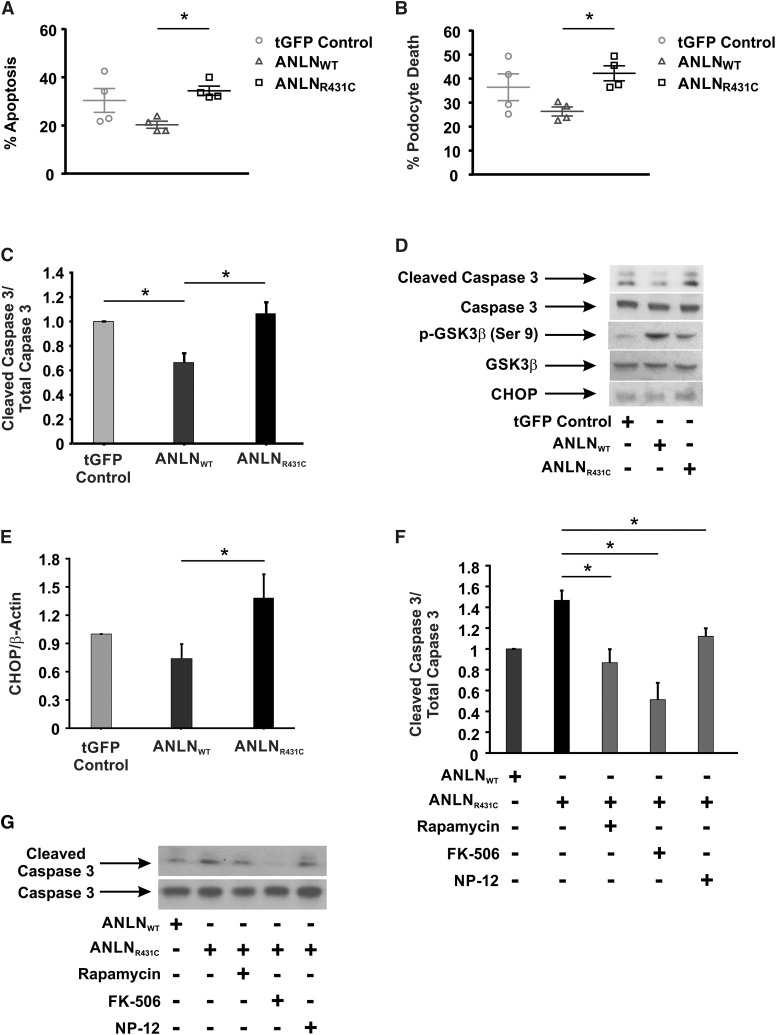
Figure 6.
Overexpression of anillinR431C (ANLNR431C) induces endoplasmic reticulum (ER) stress and apoptosis in podocytes. (A and B) Apoptosis was examined in the control and experimental podocyte cell lines after 48 hours of serum starvation using Annexin V and 7-AAD staining. FACS analysis revealed increased apoptosis and total cell death in the ANLNR431C-expressing podocytes compared with the wild type. (C and D) Cell lysates from control, ANLNWT, and ANLNR431C podocyte cell lines exposed to 48 hours of serum starvation were examined for activation of caspase 3 apoptotic signaling. Cleaved caspase expression was significantly increased in the ANLNR431C cell line compared with the ANLNWT. (D) Representative Western blots are shown for cleaved caspase 3, total caspase 3, and β-actin expression. (C) Quantification of Western blot analysis shows a decrease in caspase 3 apoptotic signaling in ANLNWT cells compared with GFP controls (P=0.03) but an increase in ANLNR431C compared with ANLNWT cells (n=4; P=0.01). (D) Representative Western blots images showing expression of cleaved caspase 3, total caspase 3, phosphoglycogen synthase kinase 3β (phospho–GSK-3β) S9, total GSK-3β, and C/EBP homologous protein (CHOP) in serum-starved turbo GFP (tGFP) control–, ANLNWT-, and ANLNR431C-expressing podocytes. (E) Quantification of CHOP expression revealed a significant increase in CHOP expression in ANLNR431C compared with ANLNWT cells (n=3; P=0.04). (F) Twenty-four–hour treatment of the serum-starved cells with 100 nM mTOR inhibitor rapamycin, 1 μM calcineurin phosphatase (Cn) inhibitor FK-506, or 2.5 μM GSK-3β inhibitor NP-12 was able to significantly reduce cleaved caspase 3 in ANLNR431C podocytes (P=0.02, P<0.01, and P=0.05, respectively) (n=3). Representative blots for each treatments as well as control ANLNWT- and ANLNR431C-expressing podocytes are depicted in G. *P value <0.05.

