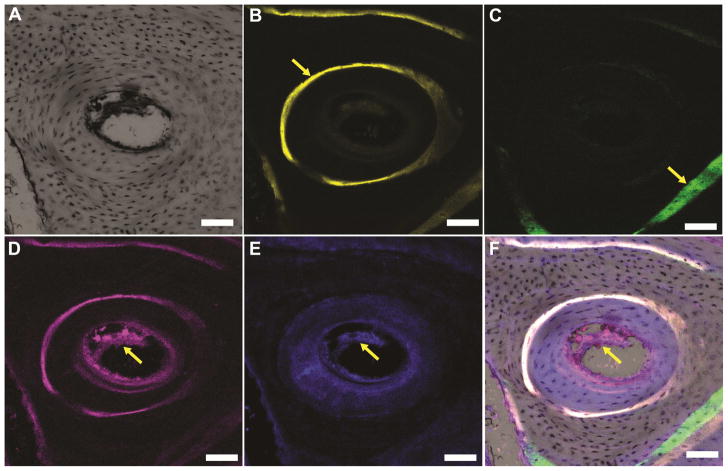
Figure 8.
High-magnification confocal microscope images (scale bar = 100 μm) of a representative Haversian system present in new bone formed near the CG/nHA-PEUR cement. (A) Bright-field image of a representative osteon. (B) Fluorescent image of the fluorochrome xylenol orange verifies that new bone was mineralizing to form Haversian systems at 8 weeks (yellow arrow). (C) Fluorescent image of the fluorochrome calcein green (yellow arrow) reveals evidence of new bone formation outside the osteon at 4 weeks. (D-E) Immunofluorescence staining of the endothelial markers (D) CD 31 (stained red, yellow arrow) and (E) endomucin (stained blue, yellow arrow) reveals the formation of Haversian systems. (F) Overlay of the images in Panels B – E.

