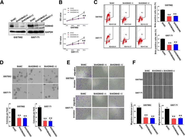
Fig. 3.
KDM4D silencing reduces GIST cell proliferation, migration and invasion . a GIST882 and GIST-T1 cells stably expressing ShNC or ShKDM4D were harvested. KDM4D protein expression was examined by Western blot, and GAPDH served as the loading control. b The viability of GIST882 and GIST-T1 cells stably expressing ShNC or ShKDM4D was detected using CCK8 assays. **p < 0.01 (c) DNA synthesis ability was examined in cells stably expressing ShNC and cells stably expressing ShKDM4D using BrdU/PI assays. Quantitative analysis of DNA synthesis is presented in the right panel. **p < 0.01 (d) Approximately 500 GIST882 and GIST-T1 cells stably expressing ShNC or ShKDM4D were seeded on 6-well plates for soft agar colony formation assays. Quantitative analysis of colony formation is presented in the lower panel. **p < 0.01 (e) Both GIST882 and GIST-T1 cells stably expressing ShNC or ShKDM4D were plated in the upper chamber with/without Matrigel for 24 h. Then, cell migration and invasion were assessed by observing the cells that migrated to the underside of the Transwell insert. f Wound healing assays were used to examined cell migration in GIST882 and GIST-T1 cells stably expressing ShNC or ShKDM4D. Quantitative analysis of migration distance is presented in the lower panel. **p < 0.01

