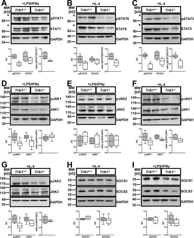
Figure 4.
Absence of Trib1 impairs JAK-mediated STAT activation. BMDMs from WT and Trib1−/− mice were treated with LPS (100 ng/ml)/IFNγ (20 ng/ml) (M1 polarization; A, D, E, and I) or IL-4 (20 ng/ml) (M2 polarization; B, C, and F–H) for 30 min. Total cell lysates were prepared and analyzed by immunoblotting. Abundance of total and phosphorylated protein was determined for STAT1 (A), STAT6 (B), STAT3 (C), JAK 1 (D and F), JAK2 (E), and JAK3 (G). Protein levels of SOCS1 and SOCS3 were determined under M2 (H) and M1 (I) polarizing conditions. An anti-GAPDH antibody was used to assess equal protein loading (A–I). All panels display a representative blot from two to four independent experiments with two samples per genotype used in each experiment. Relative quantification of phosphorylated and total protein was performed using the ImageJ software and is shown below each blot. For quantitative analysis, data were normalized to the WT group and presented as fold change (mean ± S.D.). *, p < 0.05; **, p < 0.01. The striped columns represent WT control, and the solid white columns represent Trib1−/− BMDMs. The asterisks in A–C indicates that membranes were cut below the 115-kDa marker to allow parallel incubation with different antibodies.

