Figure 6.
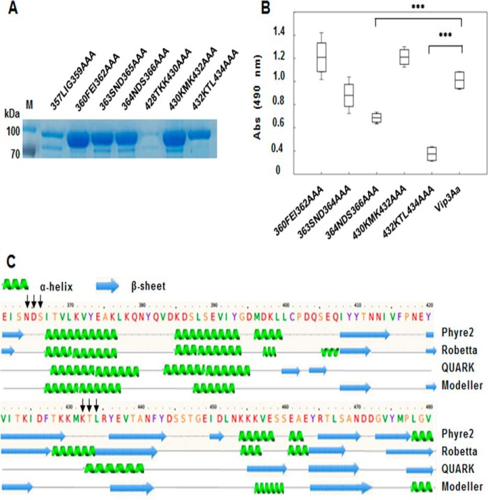
Analysis of binding of Cry9Aa to different Vip3Aa mutants. A, expression of different Vip3Aa mutants located in the identified regions. In each mutant, three selected amino acids were substituted by alanine. The expression of each Vip3Aa triple mutant was analyzed by SDS-PAGE. B, ELISA binding assays of 28 nm biotinylated Vip3Aa mutants to Cry9Aa (0.5 μg) fixed on the plate, showing that the binding was affected to Vip3Aa-N364A/D365A/S366A and Vip3Aa-K432A/T433A/L434A mutants with significant differences (***, p < 0.005) analyzed by t test. Error bars represent S.D. C, secondary structure of Vip3Aa predicted by different programs (Phyre2 (53), Robetta, QUARK (54, 55), and Modeller (56, 57)). The black arrows indicate the mutagenized sites of Vip3Aa, which are probably located in loop regions. M, molecular mass markers; Abs, absorbance.

