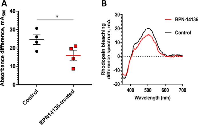
Figure 6.

Comparison of rhodopsin levels in BPN-14136–treated and control BALB/cJ mice. The amount of rhodopsin was measured spectrophotometrically in retinal extracts from BPN-14136–treated and control mice (A). Treated mice (n = 4) were orally administered BPN-14136 at 20 mg/kg for 2 weeks. The amount of pigment is expressed as absorbance difference before and after light irradiation at the wavelength of maximum absorption of rhodopsin (λmax = 500 nm). Each data point represents the average of three measurements from one retinal extract prepared from two BPN-14136-treated or control animal retinas. An unpaired t test on the ungrouped data revealed a statistically significant 25% decrease in rhodopsin concentration in treated animals compared with the control group (p < 0.05). B, representative rhodopsin bleaching difference spectra for control and BPN-14136–treated mice. The representative bleaching difference spectra are taken from a single retinal extract prepared from two mouse eyes of control and compound-treated animals.
