Figure 2.
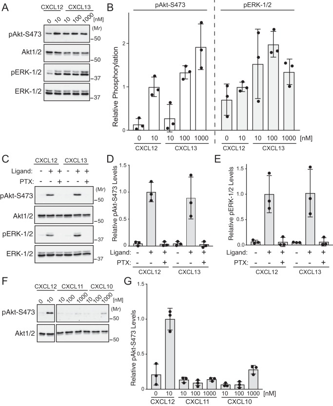
Chemokine-mediated signaling in HeLa cells. A, immunoblot analysis of CXCL13-induced phosphorylation of Akt and ERK-1/2 in HeLa cells. Serum-starved cells were treated with increasing doses of CXCL13 (10–1000 nm), 10 nm CXCL12, or vehicle for 5 min. Whole-cell lysates were analyzed by immunoblotting for the indicated proteins. Representative immunoblots from three independent experiments are shown. B, immunoblots were analyzed by densitometry. Bars represent the mean of the relative levels of pAkt-Ser473 or pERK-1/2 to CXCL12 treated samples. Error bars represent the S.D. Data were analyzed by two-way ANOVA followed by Tukey's multiple comparison test. C, CXCL13-instigated phosphorylation of Akt and ERK-1/2 is pertussis toxin–sensitive. HeLa cells were serum-starved and treated with or without 50 ng/ml pertussis toxin for 7 h followed by 10 nm CXCL12 or 100 nm CXCL13. Whole-cell lysates were analyzed by immunoblotting for the indicated proteins. Representative immunoblots from three independent experiments are shown. D and E, immunoblots were analyzed by densitometry. Bars represent the mean of the relative levels of pAkt-Ser473 (D) or pERK-1/2 (E) to CXCL12-treated samples. Error bars represent the S.D. F, the effect of chemokines CXCL11 and CXCL10 in HeLa cells on phosphorylation of Akt or ERK-1/2. Serum-starved cells were treated with vehicle or increasing doses (10–1000 nm) of CXCL11, CXCL10, or 10 nm CXCL12 for 5 min. Whole-cell lysates were analyzed by immunoblotting for the indicated proteins. Representative immunoblots from three independent experiments are shown. Panels are separated from each other to remove irrelevant intervening bands but are from the same exposure. G, immunoblots were analyzed by densitometry. Bars represent the mean of the relative levels of pAkt-Ser473 to CXCL12-treated samples. Error bars represent the S.D.

