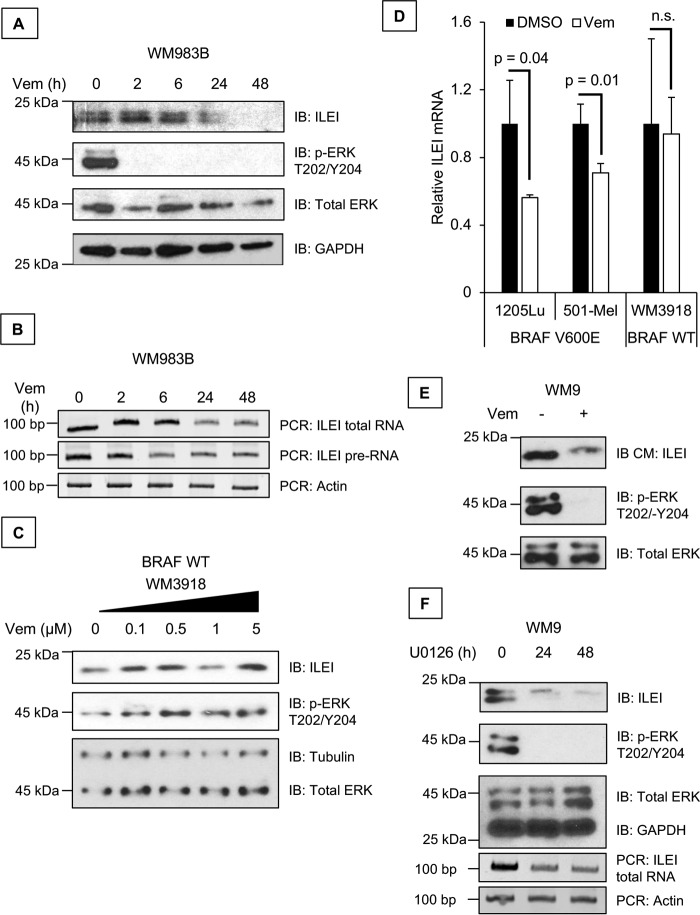
Figure 2.
Vemurafenib inhibits ILEI expression. A, immunoblot (IB) analysis of ILEI, p-ERK, ERK, and GAPDH levels in WM983B melanoma cell lines treated for 0 to 48 h with vemurafenib (1 μm). B, RT-PCR analysis of ILEI, ILEI intron indicates that PCR primers targeted the pre-RNA but not mRNA of the ILEI gene, and Actin levels in WM983B melanoma cell lines treated for 0 to 48 h with vemurafenib (1 μm). C, immunoblot analysis of ILEI, p-ERK, ERK, and Tubulin levels in WM3918 melanoma cell lines treated for 24 h with vemurafenib (0–5 μm). D, bar diagram showing quantitative RT-PCR analysis of ILEI levels in 501-Mel, 1205Lu, or WM3918 melanoma cell lines treated for 24 h with vemurafenib (1 μm). n = 3, mean ± S.D., p value indicated by Student's t test as compared with vehicle treatment, transcript values are normalized to GAPDH. E, immunoblot analysis of ILEI, p-ERK, and ERK levels in WM9 melanoma cell lines treated for 0 or 24 h with vemurafenib (1 μm). IB CM indicates that the serum-free medium condition for 24 h during vemurafenib treatment was harvested and TCA precipitated for immunoblot analysis. F, immunoblot and RT-PCR analysis of ILEI, p-ERK, ERK, GAPDH, and actin levels in WM9 melanoma cell lines treated for 0 to 48 h with U0126 (MEKi, 10 μm).

