Figure 3.
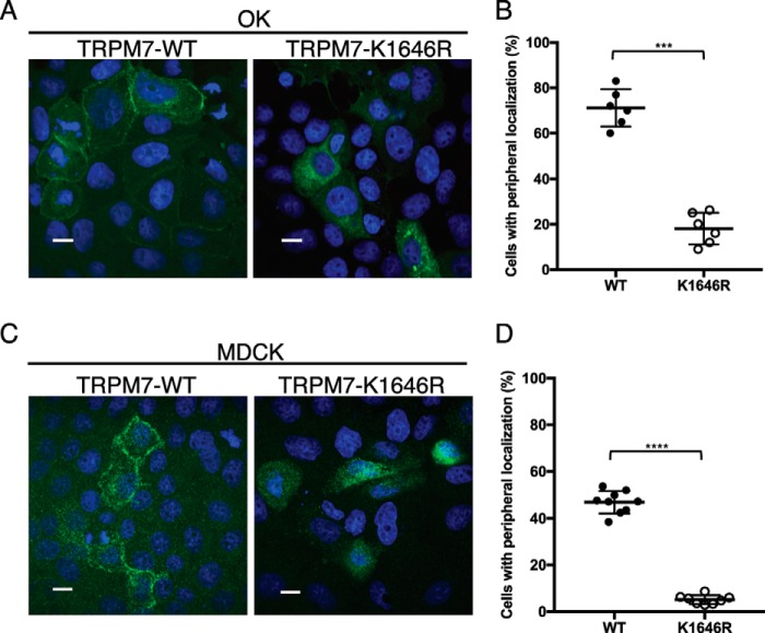
Inactivation of TRPM7 kinase affects the cellular localization of the channel in polarized epithelial cells. A and C, representative confocal images of HA–TRPM7–WT and HA–TRPM7–K1646R cellular localization in OK (A) and MDCK (C) cells after 48 h of expression. Scale bars, 10 μm. TRPM7–WT readily localizes to the lateral membranes, whereas the kinase-inactive mutant TRPM7–K1646R is retained intracellularly. B and D, quantification of the percentage of cells expressing TRPM7 localized to apical and basal–lateral sides of OK (B) and MDCK (D) cells. Each data point represents a section on the slides where 100–150 cells stained positive for TRPM7 were counted. Two to three nonoverlapping sections per slide were analyzed. Three independent experiments were performed (means ± S.D.). ***, p = 0.0001; ****, p < 0.0001.

