
Figure 3.
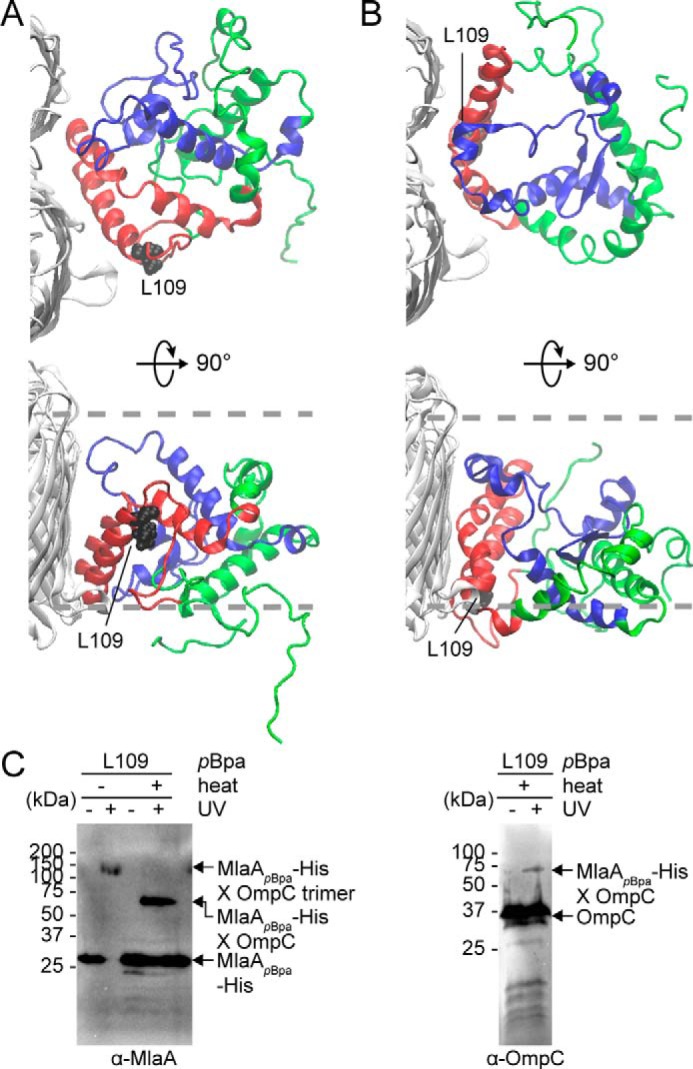
Molecular models of the OmpC–MlaA complex depict how MlaA may interact with OmpC in the OM bilayer. A and B, representative MlaA structures bound to OmpC in two possible orientations selected from all-atomistic MD simulation trajectories. MlaAD61–K124 and MlaAF133–R205 peptides are highlighted in red and blue, respectively, as in Fig. 2D. Leu109 is labeled and depicted as black spheres on MlaA. The OM boundaries are indicated as gray dashed lines. The figures were generated using the program VMD (55). C, immunoblots showing UV-dependent formation of a cross-link between MlaA and OmpC in ΔmlaA cells expressing MlaAL109pBpa-His. As expected, the cross-linked product also exhibits heat-modifiable gel shift, indicative of the presence of OmpC.