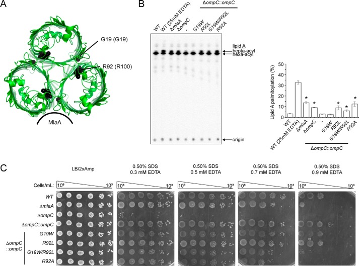
Figure 7.
A specific mutation in the dimeric interface of the OmpC trimer results in perturbation in OM lipid asymmetry. A, cartoon representation of the crystal structure of OmpC trimer illustrating the positions of Gly19 and Arg92 region (numbering in OmpF is given in parentheses). The figure was generated using the program PyMOL (56). B, analysis of SDS/EDTA sensitivity of WT and ΔompC strains producing the indicated OmpC variants from the chromosomal locus. C, representative TLC/autoradiographic analysis of 32P-labeled lipid A extracted from stationary phase cultures of strains described in B. Equal amounts of radioactive material were spotted for each sample. Average percentages of palmitoylation of lipid A and the standard deviations were quantified from triplicate experiments and plotted on the right. For Student's t tests: *, p < 0.005 compared with ΔompC::ompC.

