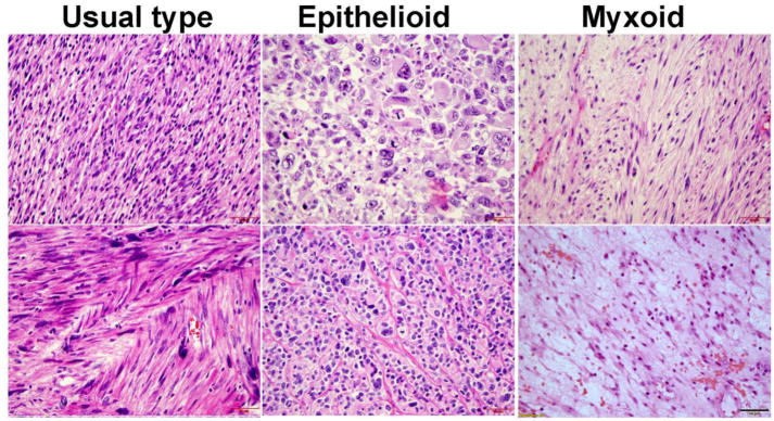
Figure 1.
Photomicrographs (40X) illustrating examples of classic LMS (left: cellular spindled cells with significant nuclear atypia and eosinophilic cytoplasm), epithelioid LMS (middle: large round cells with pleomorphic nuclei) and myxoid LMS (right: less cellular spindled cells with smaller elongated nuclei in the background of myxoid stroma).

