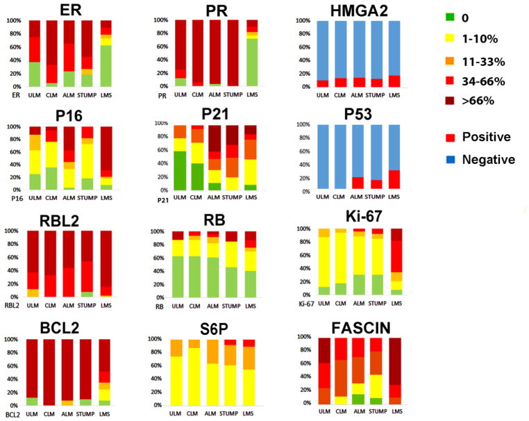
Figure 4.
Semiquantitative values for the selected markers in five different types of uterine smooth muscle tumors. Colors ranging from light green to dark red indicate the five scale cutoff ranges for of immunopercentage as listed on right. P53 and HMGA2 were shown as positive (red) and negative (blue) based on staining patterns.

