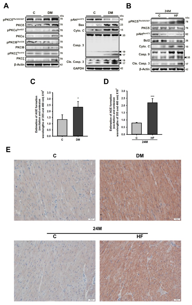
Figure 3. Cardiac PKCδ expression and phosphorylation as well as apoptosis-related proteins are increased in diseased animal models with elevated circulatory AGE levels.

Western blot analysis of the cardiac expression and phosphorylation levels of PKC isoforms and apoptosis-related proteins in rats with (A) diabetes mellitus (DM) and (B) a high-fat (HF) diet. Serum AGE levels in rats with (C) DM; (D) HF diet. These are cropped blots; full-length blots are presented in Suppl. Figure S3. Protocols for animal models with DM and HF diets and serum AGE analysis were described in the methods section. (E) Cardiac expression of phosphorylated PKCδ was examined by immunohistochemistry analysis. Bars indicate the mean ± SEM obtained from experiments performed in triplicate. *P<0.05 and ***P<0.001 compared with the control group. DM, Diabetes mellitus; HF, high-fat diet.
