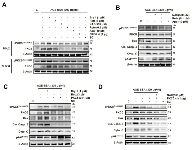
Figure 5. AGE-BSA-induced cardiomyocyte apoptosis is mediated through ROS-dependent PKCδ activation.
NRVM and H9c2 cells were exposed to AGE-BSA (300 μg/ml) for 24 h. Cells were co-treated with bryostatin 1, a PKCδ activator (100 nM), rottlerin, a PKCδ inhibitor (3 μM), NAC (500 μM), Rote (0.1 μM), Apo (10 μM) or siPKCδ (1 μg). (A) Expression and phosphorylation of PKCδ and (B, C, D) apoptosis-related proteins were examined by western blot analyses. These are cropped blots, full-length blots of PKCδ and pPKCδ are presented in Suppl. Figure S5. β-Actin was used as a loading control. N-acetylcysteine, NAC; Rote, rotenone; APO, apocynin; SC, scramble.

