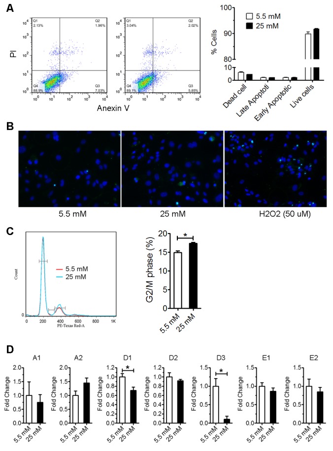
Figure 2. High glucose induced astrocyte cell cycle arrest without increasing apoptosis.

A) Flow cytometry analysis of Annexin-V and PI staining of astrocytes cultured in normal (5.5 mM) and high glucose (25 mM) medium for 3 days (n=4). B) TUNEL staining (green) of astrocytes cultured in normal (5.5 mM) and high glucose (25 mM) for 3 days. Astrocytes were treated with 50 μM H2O2 for ~12 hours as positive control. Cells were counter stained with DAPI (blue). C) Astrocytes were cultured in normal (5.5 mM) and high glucose (25 mM) for 3 days. Then astrocytes were stained with PI and analysis by flow cytometry (* p<0.05 vs 5.5 mM, n=4). D) Real-time rtPCR analysis of cyclin expression in astrocytes cultured in normal (5.5 mM) and high glucose (25 mM) medium for 2 days (* p<0.05 vs 5.5 mM, n=4).
