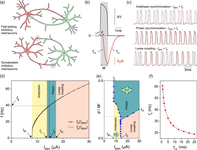
Figure 1.
Synchronization of a pair of mutually inhibitory neurons and its dependence on synaptic kinetics. (a) Fast-spiking soma-projecting and somatostatin dendrite-projecting interneurons. Synapses located on dendrites effectively delay the inhibition of the postsynaptic neuron by 0– 800 μs. (b) Inhibitory postsynaptic current (red line) evoked by a presynaptic action potential (black line) applied to a VLSI synapse. Synaptic kinetics: inhibition delay d, neurotransmitter docking time τd, undocking time τu, and spike width W. (c) Membrane voltage oscillations of mutually inhibitory neurons below, at, and above the synchronization current, Is. τu = 1.5 ms. (d) Frequency-current dependence of a VLSI neuron (square symbols) and frequency-current dependence of phase-locked oscillations (red line). Their intercept gives the frequency (fs) and current (Is) of phasic oscillations. Domains of synchronized oscillations at d = 0.2W (vertical bands). (e) Phase diagram of synchronization in the d − Istim plane where delay d is normalised by the spike width W. Two alternative mechanisms contribute to synchronization in local inhibitory networks: a change in current stimulation (PVB: parvalbumin neuron-type synchronization) and an increase in inhibition delay (SST: somatostatin neuron-type synchronization). (f) Frequency of phasic oscillations as a function of the decay time of the postsynaptic current.

