Fig. 3.
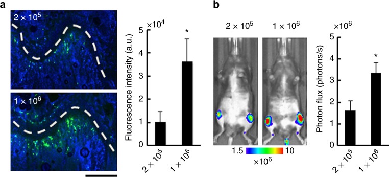
Injection of larger number of cancer cells enhances bone metastasis development. a Fluorescence microscopic analysis of cancer cells in bone marrow of the femur. Representative fluorescence images of the femur (left) and fluorescent intensities of cancer cells (right) at 30 min after CA injection of green-fluorescently labeled LLC/luc cells (2.0 × 10⁵ or 1.0 × 10⁶ cells). Cells were stained with Hoechst dyes (blue). White-dashed lines indicate growth plates. A scar bar is 200 μm. n = 4, *P < 0.05 (two-side student’s t-test). b Development of bone metastasis after injection of different number of LLC/luc cells. Representative BL images (left) and BL intensity in hind limbs (right) at 14 days after CA injection of LLC/luc cells (2.0 × 10⁵ or 1.0 × 10⁶ cells). n = 8, *P < 0.05 (two-side student’s t-test). Error bars indicate s.e.m.

