Figure 7.
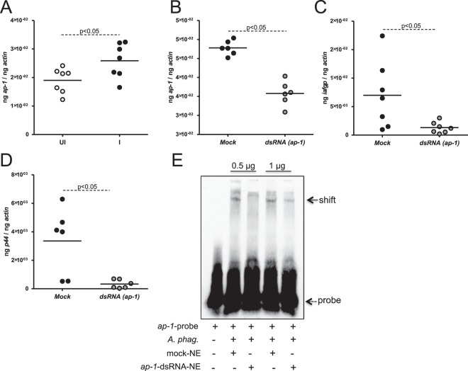
Knockdown of ap-1 affects expression of iafgp gene and A. phagocytophilum burden in ISE6 cells in vitro. (A) QRT-PCR results showing ap-1 transcripts in uninfected or A. phagocytophilum-infected ISE6 cells at 48 hrs p.i. The QRT-PCR results for the expression of ap-1 (B) or iafgp (C) transcripts in tick cells upon treatment with mock or ap-1-dsRNA is shown. (D) Bacterial burden in mock or ap-1-silenced ISE6 cells at 24 hrs p.i. is shown. (E) Gel shift assays with biotinylated iafgp-promoter probe containing AP-1 binding site (DS704943, 18144–18193 bp) and nuclear proteins (0.5, 1 µg) prepared from A. phagocytophilum-infected mock or ap-1-silenced ISE6 cells is shown. Dotted arrow indicates free probe and solid arrow indicates shift. Each circle in panels A–D indicates RNA/DNA samples generated from one tick cells culture well. Statistical analysis was performed using Student’s t test and P value less than 0.05 was considered significant. NE indicates nuclear extracts and + or − indicates presence or absence, respectively.

