Figure 5.
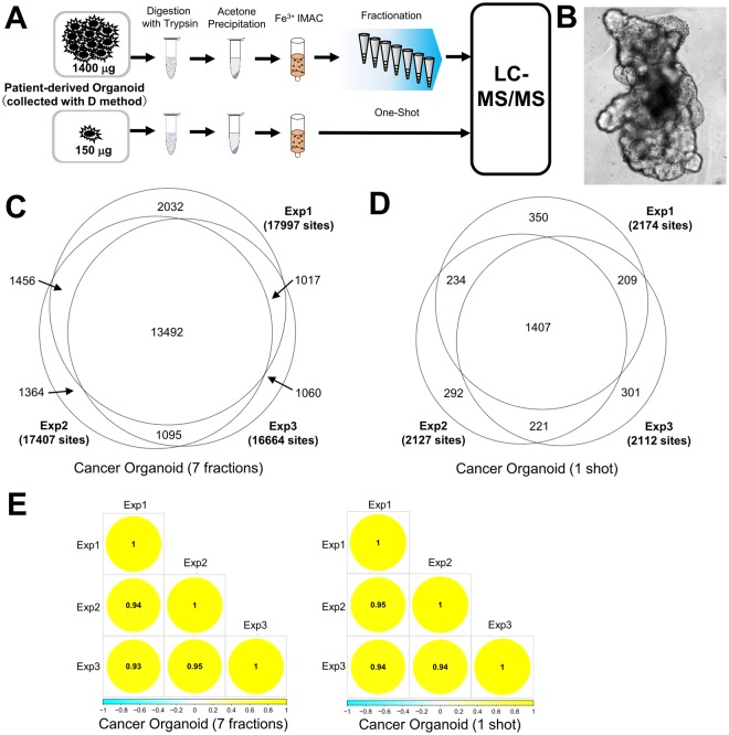
Large-scale and small-scale phosphoproteomics using patient-derived organoids. (A) Workflow of large-scale and small-scale phosphoproteomics. (B) Picture of a patient-derived organoid. (C) Proportional Venn diagram of class 1 phosphosites identified from patient-derived organoids with fractionation. (D) Proportional Venn diagram of class 1 phosphosites identified from patient-derived organoids in a one-shot analysis. (E) Correlation matrix of phosphosites quantified in all triplicate fractionated and one-shot phosphoproteomics experiments.

