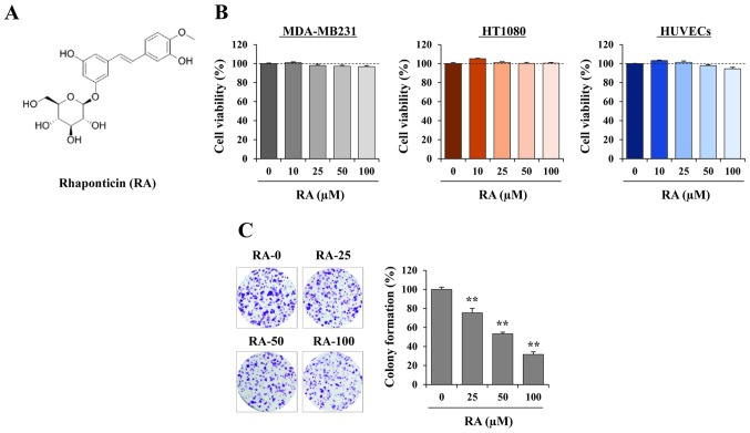
Figure 1.
RA exhibits no cytotoxicity and suppresses colony-forming activity in MDA-MB231 cells. (A) Chemical structure of RA. (B) MDA-MB231 cells, HT1080 cells and HUVECs were seeded into 96-well culture plates in triplicate and treated with the indicated concentrations of RA. At 48 h after treatment, the proportion of viable cells was determined using the Cell Counting Kit-8 assay, then compared with that of RA-untreated cells, and expressed as the mean ± SD. (C) Anchorage-dependent colony formation of MDA-MB231 cells in the presence or absence of RA was determined by counting visible colonies following staining with crystal violet solution (n=3 per group). The relative values are expressed as the mean ± SD. **P<0.01 vs. untreated control. RA, rhaponticin; HUVEC, human umbilical vein endothelial cell; SD, standard deviation.

