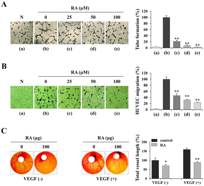
Figure 3.
RA suppresses the angiogenic activity of HUVECs. (A) HUVECs were treated with or without the indicated concentrations of RA for 24 h, suspended in EGM-2, and seeded in basement membrane extract-coated wells. Following incubation for 4 h, capillary-like tube formation was measured. RA-untreated HUVECs in EBM-2 were used as a negative control. The number of tubes in triplicate samples was counted. (B) RA-treated or untreated HUVECs were suspended in EBM-2 and seeded into the upper chamber of the Transwell. The EGM-2-induced migration of HUVECs for 12 h was observed following staining with crystal violet solution (n=5 per group). (C) On ED day 6, RA (100 µg) and VEGF (200 ng) in PBS (20 µl) were loaded onto filter discs and then carefully placed on the CAM. Following resealing the windows with adhesive tape, the eggs were incubated for 3 days. On ED day 9, the vasculature in the eggs was photographed and the total vessel length (n=3 per group) was measured by ImageJ software. The data are representative of three independent experiments and expressed as the mean ± SD. *P<0.05 and **P<0.01 vs. RA-untreated control. RA, rhaponticin; HUVEC, human umbilical vein endothelial cell; EGM-2, endothelial cell growth medium-2; EBM-2, endothelial cell basal medium-2; N, negative control; ED, embryonic development; VEGF, vascular endothelial growth factor.

