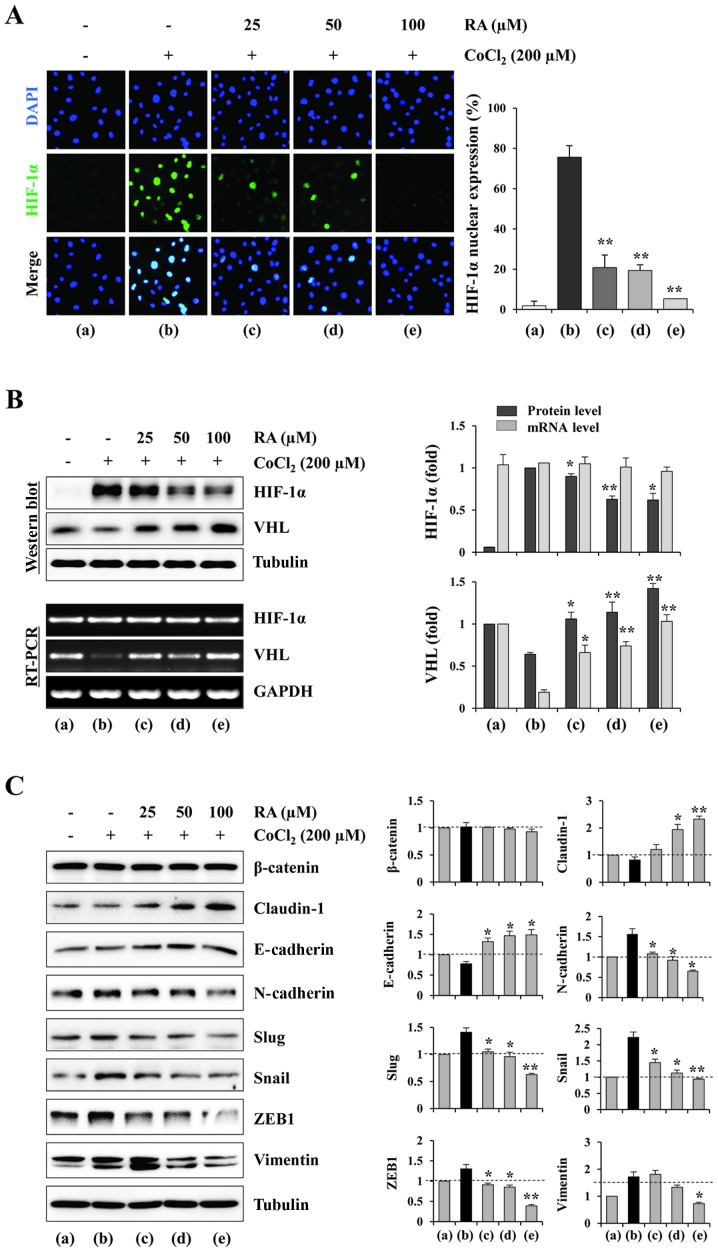
Figure 5.
RA inhibits the nuclear expression and accumulation of HIF-1α, and regulates EMT-associated proteins in HT1080 cells under hypoxic conditions. (A) Cells grown in glass-bottomed dishes were treated with RA for 12 h and then stimulated with CoCl2 (200 µM) to mimic hypoxic conditions. After 6 h, nuclear HIF-1α was visualized using fluorescence immunocytochemistry. DAPI was used for counterstaining nuclei. Results are presented as the mean ± SD of five selected fields per sample, and are representative of three independent experiments. (B) The cells were incubated with or without RA for 12 h and then treated with CoCl2 (200 µM) for 3 or 6 h. The mRNA and protein levels of HIF-1α and VHL were detected using RT-PCR and western blotting, respectively. The relative band intensities were quantitated using ImageJ software following normalization to GAPDH and tubulin expression, respectively. (C) The cells were treated with or without RA for 12 h and then stimulated with CoCl2 (200 µM) for 24 h. Cell lysates were subjected to western blotting against endothelial-mesenchymal transition-associated proteins. The relative band intensities were calculated using ImageJ software following normalization to tubulin expression. Results are expressed as the mean ± SD of two independent experiments. *P<0.05 and **P<0.01 vs. RA-untreated control. RA, rhaponticin; HIF, hypoxia-inducible factor; SD, standard deviation; VHL, von Hippel-Lindau protein; RT-PCR, reverse transcription-polymerase chain reaction; E-cadherin, epithelial cadherin; N-cadherin, neuronal cadherin; ZEB1, zinc finger E-box-binding homeobox 1.

