Figure 2.
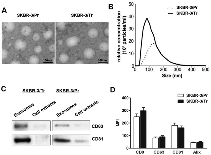
Characterization of exosomes released from trastuzumab-resistant and -sensitive SKBR-3 cells. (A) Transmission electron microscopy images of the exosomes released by SKBR-3/Pr and SKBR-3/Tr cells. (B) Nanoparticle tracking analysis on an LM10 Nanosight unit demonstrating a mean size of 100 nm for SKBR-3/Tr and 120 nm for SKBR-3/Pr exosomes. The size distribution and relative concentration were calculated using the Nano-sight software. (C) Exosomal protein marker (CD63 and CD81) detection by western blotting from purified exosomes and cell extracts. (D) Flow cytometric analysis of the MFI for a panel of exosomal markers: CD9, CD63, CD81 and Alix. Data are presented as the median ± interquartile range of triplicate experiments. MFI, mean fluorescence intensity; CD, cluster of differentiation; Alix, programmed cell death 6-interacting protein.

