Figure 3.
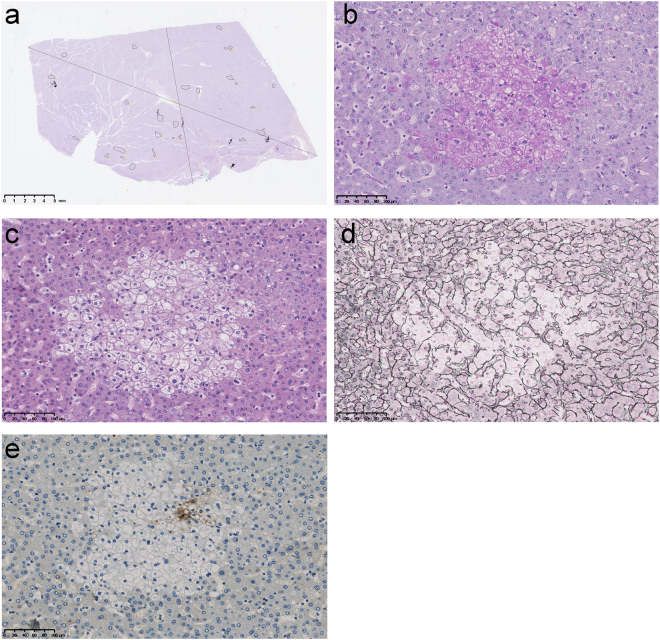
Illustration of the hepatic foci of cellular alteration observed in non-tumor liver parenchyma of Peruvian patients with hepatocellular carcinoma. (a) NTL section from a 37-year-old Peruvian male individual with a 16-cm-diameter well differentiated (G1), trabecular growth pattern HCC. The surrounded areas indicate location and size of liver clear cell foci within the section. NTL section dimension; 25.5 mm-length, 15.1 mm-width. Scale bar; 5 mm. (b–e) Representative features in a series of serial NTL sections from a 32-year-old Peruvian male individual with a 14-cm-diameter moderately differentiated (G2), trabecular growth pattern HCC under medium power magnification (30x). Scale bars; 100 µm. (b) A hepatic focus of cellular alteration under PAS staining. This histological feature was significantly associated with Patient Group by OPLS-DA (Fig. 2b). (c) The same focus than in (b) under hematoxylin–eosin staining. The focus is displaying a clear cell-like appearance. (d) The same focus than in (b,c) under Gömöri reticulin staining. It is observed a distortion of the trabecular architecture over the focal area. (e) GS immunohistochemistry on the same focus than in (b–d). GS expression is observed heterogeneously in some cells of the hepatic focus of cellular alteration.

