Figure 4.
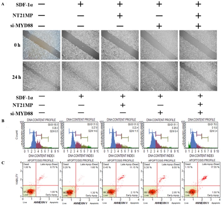
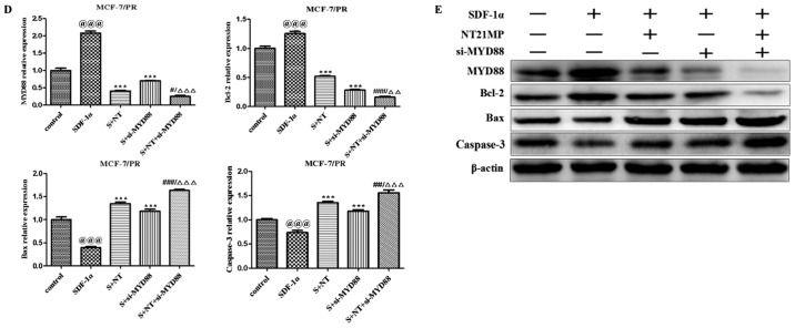
Biological effects of NT21MP and si-MYD88 on PR cells. (A) Effects of NT21MP and si-MYD88 on cell migration and invasion were measured using a wound-healing assay. (B) Effects of NT21MP and si-MYD88 on cell cycle were analyzed using PI staining and flow cytometry. (C) Effects of NT21MP and si-MYD88 on cell apoptosis were evaluated using Annexin V/PI staining and flow cytometry. (D) Reverse transcription-quantitative polymerase chain reaction analysis was used to detect the effect of NT21MP and si-MYD88 on the mRNA levels of target genes, cell cycle, and apoptosis-related factors. (E) Western blot analysis results of the protein level of target gene, cell cycle, and apoptosis-related factors in PR cells were consistent with the mRNA results. The data are presented as the mean ± standard deviation of three independent experiments. @@@P<0.001, compared with the control group; ***P<0.001, compared with SDF-1α treatment; #P<0.05, ##P<0.01 and ###P<0.001, compared with S + NT21MP treatment; ∆∆P<0.01 and ∆∆∆P<0.001, compared with S + si-MYD88 treatment. NT21MP; 21-residue peptide derived from viral macrophage inflammatory protein II; S/SDF-1α, stromal cell-derived factor-1α; NT, NT21MP; PR, paclitaxel-resistant; MYD88, myeloid differentiation primary response gene 88; Bcl-2, B-cell lymphoma 2; Bax, Bcl-2-associated X protein; PI, propidium iodide; si, small interfering RNA.


