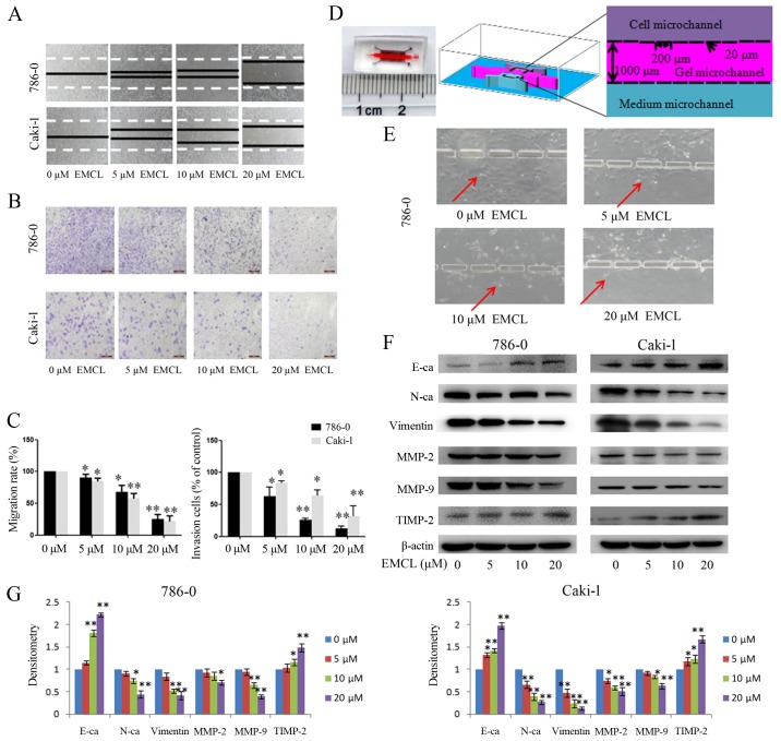
Figure 3.
Effects of treatment with EMCL on cell migration and invasion. (A) Migration of 786-0 and Caki-1 cells was analyzed using a scratch assay. Following24 h of treatment with EMCL at the indicated doses, the wound gap was observed and images were captured. (B) Cell invasion was analyzed in 786-0 and Caki-1 cells treated with EMCL at the indicated doses for 24 h. Cell invasion was observed and images were captured. (C) Percentages of migrated cells were calculated relative to the original gap and the percentage of invaded cells was calculated. (D) Image and illustration of the homemade microfluidic chip. The chip comprised two-sided micro-channels sandwiching a gel micro-channel with two rows of micro-gaps. The gel micro-channel was filled with Matrigel (pink), and the cell micro-channel and medium micro-channel were used for plating the cells (purple) and the chemoattractant (20% FBS; green), respectively. (E) Invasion of 786-0 cells to the gel micro-channel following 24 h of treatment with EMCL at the indicated doses. Red arrows indicate invasion of 786-0 cells. (F) Protein expression levels of MMP-2/9, TIMP-2, E-cadherin, N-cadherin and vimentin were analyzed by western blot analysis. (G) Quantitative analysis of proteins. Data are presented as the mean ± standard deviation of three independent experiments. *P<0.05 and **P<0.01. vs. dimethyl sulfoxide-treated group. EMCL, epoxymicheliolide; E-ca, E-cadherin; N-ca, N-cadherin; MMP, matrix metalloproteinase; TIMP-2, tissue inhibitor of metalloproteinase 2.

