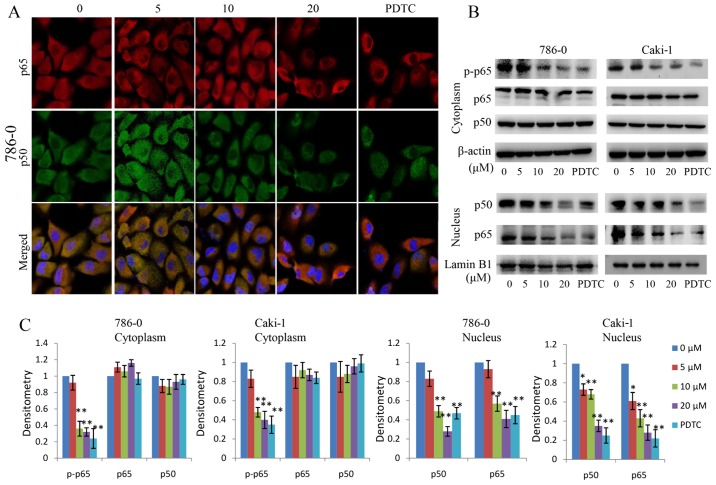
Figure 7.
Effect of EMCL on nuclear factor-κB signaling. (A) Following treatment with EMCL at the indicated doses and PDTC (30 µM) for 24 h, the subcellular localizations of p50 and p65 in 786-0 cells were examined by confocal microscopy analysis(magnification, ×630). (B) Following treatment with EMCL at the indicated doses and PDTC (30 µM) for 24 h, cytoplasmic and nuclear extracts were prepared for the western blot analysis of p-p65, p65, and p50. (C) Quantitative analysis of the proteins. Data are presented as the mean ± standard deviation of three independent experiments.*P<0.05 and **P<0.01, vs. dimethyl sulfoxide-treated group. EMCL, epoxymicheliolide; PDTC, ammonium pyrrolidine dithiocarbamate; p-, phosphorylated.

