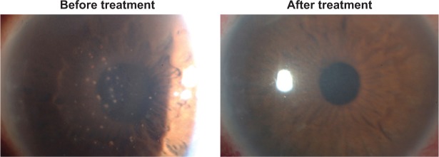
Figure 1.
Slit-lamp microscopic photograph with cytomegalovirus anterior uveitis in Patient 1 showing numerous mutton fat keratic precipitations on the central to inferior corneal endothelium with mild stromal edema and elevation of intraocular pressure.
Notes: The anterior chamber contained 2+ cells according to the Standardization of Uveitis Nomenclature grading system. After antiviral treatment, these keratic precipitations and anterior chamber cells were resolved. Additionally, corneal transparency was restored.

