
Figure 3.
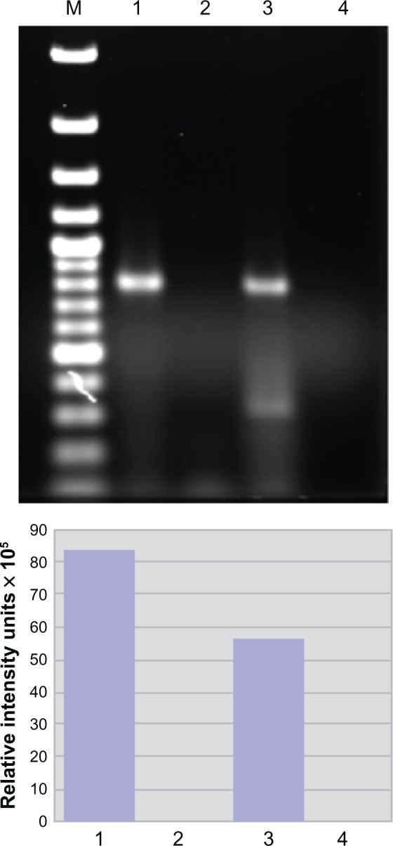
Spastin mRNA is bound to heterogeneous nuclear ribonucleoprotein (hnRNP) A1 in neuronal cells. Upper panel: agarose gel. Compared to the neuronal lysate without immunoprecipitation or input (lane 3), there is an enriched spastin mRNA signal following immunoprecipitation with anti-hnRNP A1 mouse monoclonal antibodies (lane 1). In contrast, spastin mRNA was not isolated following immunoprecipitation with a nonspecific control antibody – mouse IgG (lane 2). Lane 4 used spastin primers without lysate (control for DNA contamination). Lower panel: the image was analyzed using ImageQuant software, which provided relative fluorescent intensity of the bands.