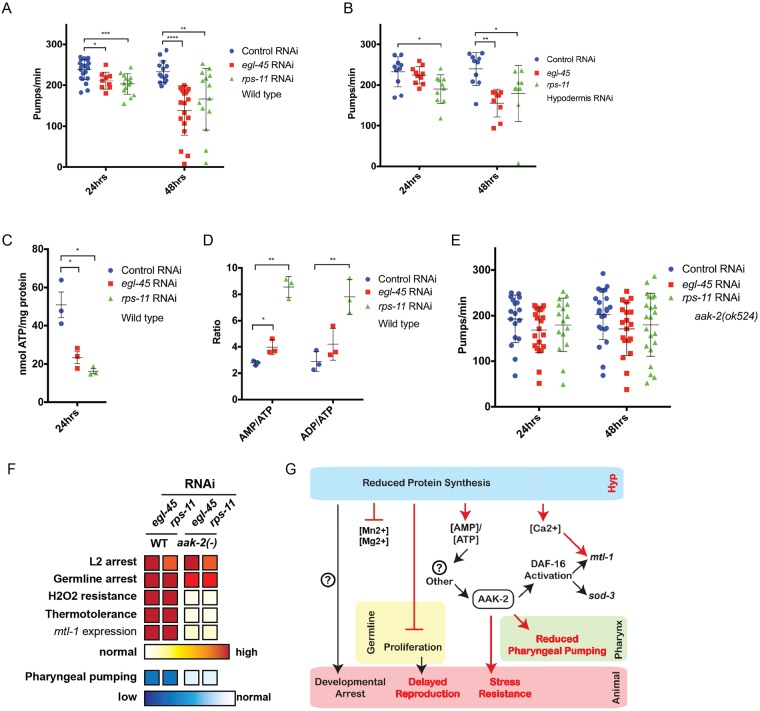
Fig 4. Protein synthesis inhibition drives a reduction in pharyngeal pumping and stress resistance through AMPK signaling.
A. Protein synthesis inhibition decreases pharyngeal pumping rates in wildtype animals in a time-dependent manner (N = 9–23 from 2–3 biological replicates). B. Reducing protein synthesis in the hypodermis is sufficient to reduce pharyngeal pumping rate (N = 9–10 from 2 biological replicates). C-D. Protein synthesis inhibition arrested larvae have reduced levels of cellular ATP, and their ratio of AMP and ADP to ATP is higher (3 biological replicates). E-F. aak-2/AMPK mutant animals fail to reduce pharyngeal pumping in response to protein synthesis inhibition (E) and although arrested, are not resistant to environmental stress (F) (N = 16–24 from 2–3 biological replicates; intensity of red and blue coloring indicates increased and decreased responses, respectively, as compared to control RNAi treated animals). G. Schematic diagram of the cell autonomous and cell non-autonomous pathways that mediate organism-level responses to protein synthesis inhibition—red colors identify findings of this study. Additional mediators (?) are likely to exist, including the genetic regulators of the developmental arrest (left). * p<0.025, ** p<0.005, *** p<0.0005, **** p<0.00005 (A-B, E: One-way ANOVA, C-D: Student's t test). See also S1 Table.

