Figure 3.
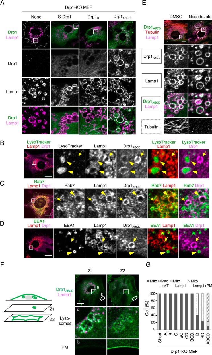
Drp1ABCD is associated with lysosomes, late endosomes, and the plasma membrane. A, we analyzed Drp1-KO MEFs that carry each Drp1 isoform using immunofluorescence microscopy with antibodies to Drp1 and Lamp1, a membrane protein that is associated with lysosomes and late endosomes. Images of Drp1-KO MEFs carrying no Drp1, S-Drp1, Drp1D, or Drp1ABCD are shown. Boxed regions are enlarged. Scale bar = 20 μm. B, Drp1-KO MEFs expressing Drp1ABCD were stained with 75 nm LysoTracker (Invitrogen, L7528) for 30 min and then subjected to immunofluorescence microscopy with antibodies against Lamp1 and Drp1. The boxed region is enlarged. Scale bar = 20 μm. C and D, immunofluorescence microscopy of Drp1-KO MEFs expressing Drp1ABCD was performed using antibodies against Rab7, Lamp1, and Drp1 (C) and EEA1, Lamp1, and Drp1 (D). Scale bars = 20 μm. E, Drp1-KO MEFs expressing Drp1ABCD were treated with 20 μm nocodazole for 4 h and subjected to immunofluorescence microscopy with antibodies to tubulin, Drp1, and Lamp1. Scale bar = 20 μm. F, confocal immunofluorescence microscopy images of Drp1-KO MEFs expressing Drp1ABCD with antibodies against Lamp1 and Drp1. Images were taken at two different focal planes, Z1 and Z2. The Z1 and Z2 focal planes are 0.9 μm apart. Boxed regions are enlarged. Scale bar = 20 μm. G, quantification of Drp1 localization. 30 cells were analyzed for each isoform. Mito, mitochondria; MT, microtubules; Lamp1, Lamp1-positive vesicles; PM, plasma membrane.

