
Figure 4.
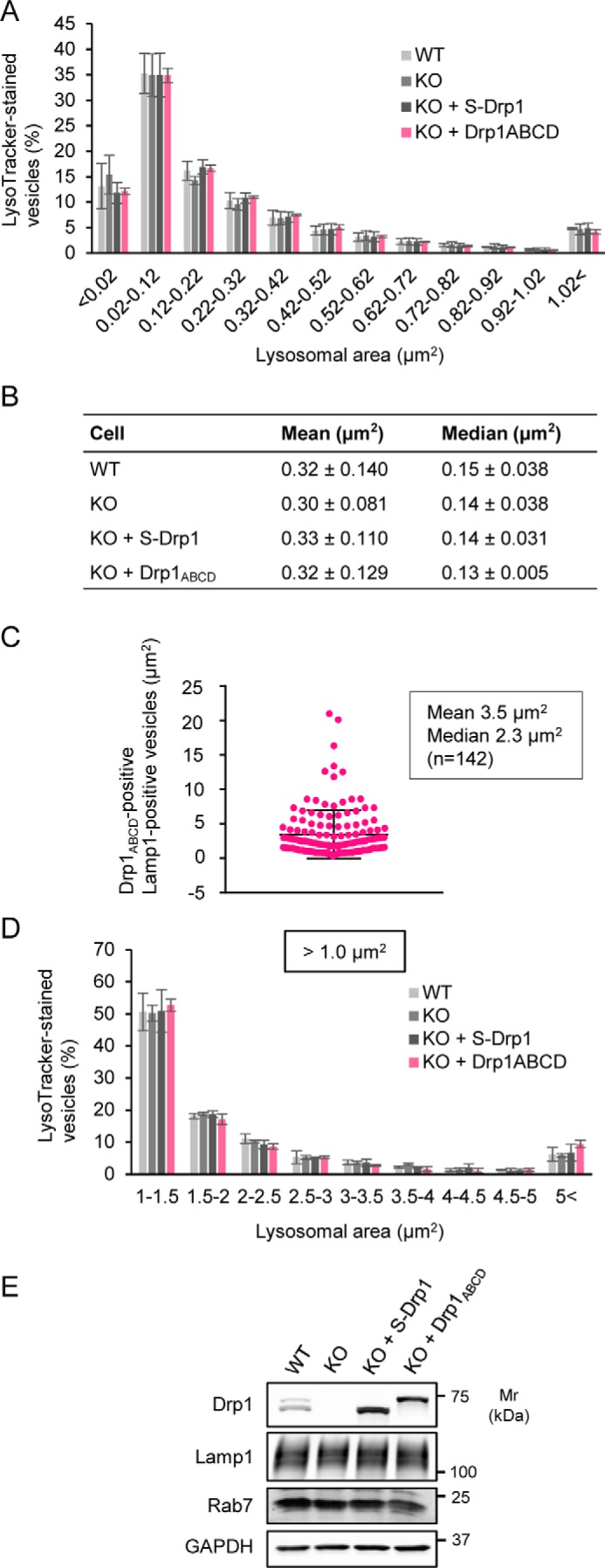
Drp1ABCD does not affect the size of lysosomes/late endosomes. A, WT MEFs, Drp1-KO MEFs, and Drp1-KO MEFs expressing either S-Drp1 or Drp1ABCD were stained with LysoTracker (75 nm for 30 min). The size distribution of the areas of LysoTracker-stained vesicles is shown. Thirty cells were analyzed. Values represent means ± S.D. (n = 3). B, the mean and median of the area of LysoTracker-stained vesicles (n = 3, 30 cells were analyzed for each cell type in each experiment). C, Drp1-KO MEFs expressing Drp1ABCD were analyzed by immunofluorescence microscopy with antibodies against Drp1 and Lamp1. The size distribution of Lamp1-positive vesicles associated with Drp1ABCD is shown. Error bars represent means ± S.D., and 142 vesicles were analyzed. D, the size distribution of LysoTracker-stained vesicles of an area larger than 1 μm2. The same set of MEFs as described in A was analyzed. Values represent means ± S.D. (n = 3). E, Western blotting of whole-cell lysates from MEFs, Drp1-KO MEFs, and Drp1-KO MEFs expressing either S-Drp1 or Drp1ABCD.