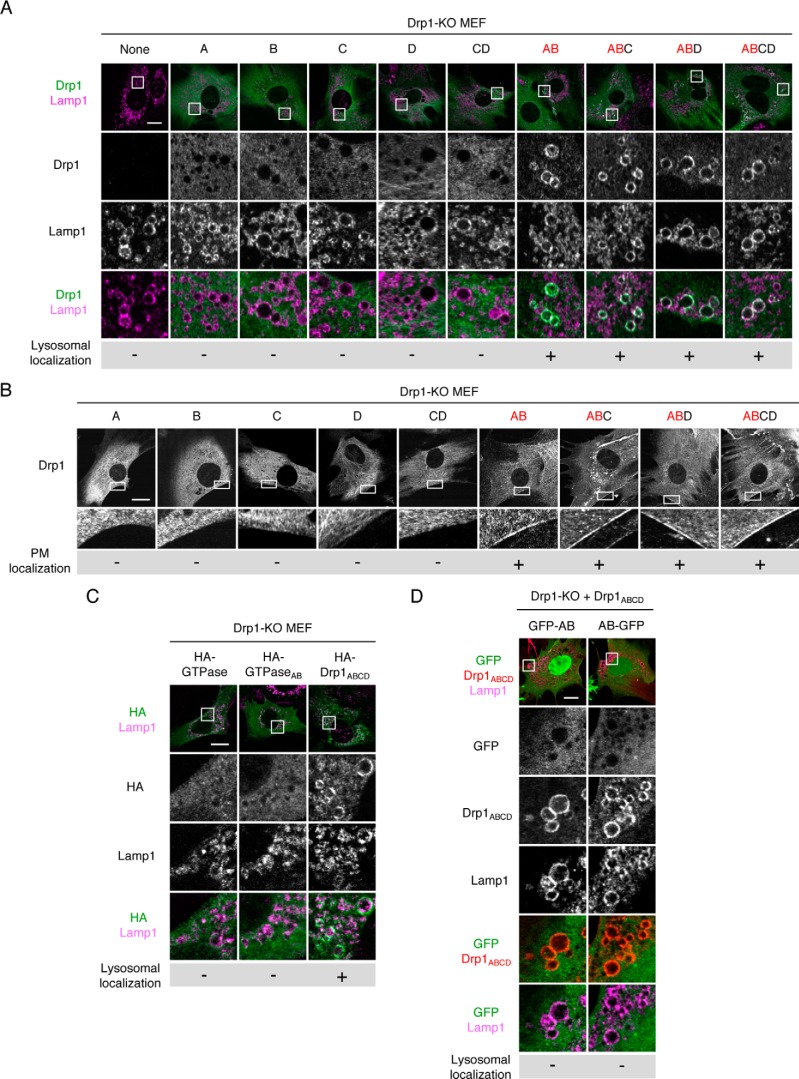
Figure 6.
Exons A and B are important for the localization of Drp1ABCD at lysosomes/late endosomes and the plasma membrane. A, Drp1-KO MEFs individually expressing the indicated Drp1 isoforms were analyzed by confocal immunofluorescence microscopy with antibodies to Drp1 and Lamp1. Scale bar = 20 μm. B, the indicated MEFs with specific Drp1 isoforms were analyzed by immunofluorescence microscopy using anti-Drp1 antibodies. Boxed regions are enlarged. Scale bar = 20 μm. C, Drp1-KO MEFs expressing the HA-tagged GTPase domain carrying no exon, AB exons, or full-length Drp1ABCD were analyzed by immunofluorescence microscopy using antibodies to the HA epitope and Lamp1. D, Drp1-KO MEFs expressing Drp1ABCD along with GFP-tagged AB exons (GFP-AB or AB-GFP) were analyzed by immunofluorescence microscopy using antibodies to Drp1 and Lamp1. Scale bar = 20 μm.

