Figure 2.
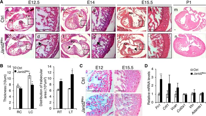
Cardiac defects were observed in Jarid2Nkx embryos. A, H&E staining was performed on transverse sections at E12.5 (a–d), E14 (e–h), E15.5 (i–l), and P1 (m and n) of Jarid2Nkx (b, d, f, h, j, l, and n) versus control (a, c, e, g, i, k, and m) mice. The boxed regions of a, b, e, f, I, and j are magnified in c, d, g, h, k, and l, respectively. Representative images of Jarid2Nkx embryos show VSD (*, f, j, and n), thin myocardium (dashed line, h and l), and disorganized hypertrabeculae (arrowheads, f and j) are shown. Arrows (d and h) indicate the increased distance between the endocardium and myocardium in Jarid2Nkx. The dotted lines separate the compact and trabecular layers. Scale bar, 100 μm. B, compact layer thickness was measured by drawing lines, and distribution of trabecular area was measured using NIH ImageJ software on the right (R) or left (L) compact layer (C) and trabecular layer (T) at E15.5. Three slides per heart were measured, n = 4. C, Alcian blue staining showed increased mucopolysaccharides in Jarid2Nkx at E12 and E15.5. The sections were counterstained with nuclear fast red. Scale bar, 100 μm. D, qRT-PCR was performed to determine the expression levels of extracellular matrix components and, a metalloproteinase, Adamts1 on control or Jarid2Nkx hearts at E13.5. The expression levels were normalized to the control; n = 3 (*, p ≤ 0.05; **, p ≤ 0.01).

