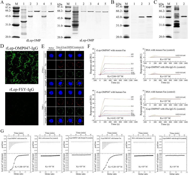
Fig. 3. Fas-binding ability of rLep-OMP047 and distribution and relative abundance of Lep-OMP047.
a Mouse or human Fas-captured proteins from dLep-OMP, detected by co-precipitation assay. Lane M: protein marker. Lane 1: dLep-OMP or aLep-OMP. Lane 2 or 4: mouse or human Fas-Fc chimera released from protein-A-coated beads and the chimera-captured proteins from dLep-OMP but not from aLep-OMP. Lane 3 or 5: mouse or human Fas-Fc chimeric protein as the control. b Lep-OMP047 distribution in a- and dLep-OMP, determined by western blot. Lane M: protein marker. Lanes 1: no rLep-OMP-IgG-binding bands in aLep-OMP. Lane 2: the single rLep-OMP-IgG-binding band in dLep-OMP. Lane 3: rLep-OMP047 control. c Relative abundance of Lep-OMP047 in dLep-OMP, assessed by western blot. Lane M: protein marker. Lane 1: the dLep-OMP-IgG-binding bands in dLep-OMP. Lane 2: rLep-OMP047 control. d Location of Lep-OMP047 on the surface of L. interrogans strain Lai, determined by confocal microscopy. The green fluorescence indicates the leptospiral surface-located Lep-OMP047. e Co-localization of Lep-OMP047 with Fas protein in cytomembrane of J774A.1 and THP-1 macrophages, determined by confocal microscopy. The red spots indicate Fas of macrophages, and the green spots indicate Lep-OMP047 attached to the cytomembrane of macrophages. The yellow spots indicate the Lep-OMP047-Fas co-localization. f Mouse or human Fas-binding ability of rLep-OMP047, determined by SPR measurement. BSA and CM5 array-linked rMs- or rHu-IgG-Fc were used as the controls. g Mouse or human Fas-binding ability of rLep-OMP047, determined by ITC detection. BSA and sample pool-loaded rMs- or rHu-IgG-Fc were used as the controls

