Figure 8.
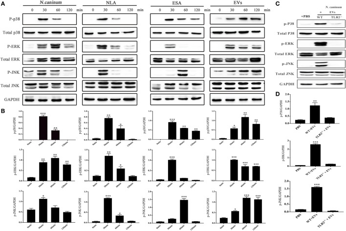
Neospora caninum extracellular vesicles (EVs) induce the phosphorylation of P38, ERK, and JNK via toll-like receptor 2 (TLR2). (A) Wild-type (WT) mouse bone marrow-derived macrophages (BMDMs) (3 × 10⁶ cells) were stimulated with N. caninum tachyzoites (MOI = 3:1, parasite:cell) or 50 µg/ml N. caninum-derived polypeptides [Neospora caninum lysate antigen (NLA), excretory secretory antigen (ESA), or EVs] for the indicated times (0, 30, 60, and 120 min) as indicated, then the phosphorylation levels of P38, ERK, and JNK were determined by immunoblot analysis. (B) Relative levels of the signals from the western blot in panel (A). (C) WT and TLR2−/− mouse BMDMs (3 × 10⁶ cells) were stimulated with 50 µg/ml N. caninum EVs for 60 min, and then the p-P38, p-ERK, and p-JNK expression were determined by immunoblot analysis. (D) Relative levels of the signals from the western blot in panel (C). The phosphorylated forms of these proteins were clearly observed at 30 min after treatment. The data are expressed as the mean ± SEM from three separate experiments, *P < 0.05; **P < 0.01; ***P < 0.001 for N. caninum EVs group versus the PBS groups.

