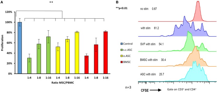
Figure 3.
Ratios ranging from 4:1 to 16:1 between responder cells [peripheral blood mononuclear cells (PBMCs)] and mesenchymal stem cell (MSC) were added to proliferation assays (n = 3 from three different human donors) stimulated with phytohemagglutinin (PHA) (A). All three types of MSC show a dose depended reduction responder cell proliferation detected by H3 thymidine assay, with the most effective inhibition by adipose-derived mesenchymal stem cell (ASC). Panel (B) shows representative results of a co-culture stimulated with PHA, gated on CD3+ and CD4+ cells confirming potent inhibition by ASC. Addition of stromal vascular fraction (SVF) cells at the same ratio resulted in a far weaker inhibition compared with cultured cells (P3).

