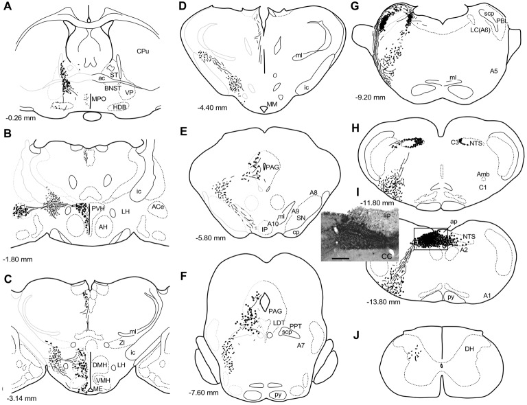
Figure 1.
A series of transverse sections of the brain from anterior (A) to posterior (J: cervical spinal cord) depicting anterogradely labeled efferent axon fibers and boutons following biotin dextran amine (BDA) injection into the NTS (H,I). The BDA injection region is indicated in the photograph (I, the caudal NTS). Fine lines and dots depict axonal fibers traveling in various directions. Large dots represent aggregations of axon boutons. The section figures and values of distance from the bregma are adopted with a slight modification from the atlas of Paxinos and Watson (1998). Aggregates of axon boutons were conspicuous in several discrete brain regions BNST in (A); central nucleus of the amygdala (ACe) and paraventricular hypothalamic nucleus (PVH) in (B); DMH and VMH in (C,D); PAG in (E,F); locus coeruleus (LC) and PBL in (G), particularly in the (PBL in G); photographs are shown in Figure 2. Note that the two ascending axon bundles (the medial forebrain bundle (MFB) and dorsal longitudinal fascicle (DLF)) appear to segregate at the ponto-midbrain junction (F,G). See also (A–I) in Figure 9. Bar in (I) inset = 200 μm. For abbreviations, see the abbreviation list. Case 1a #1.

