Figure 3.
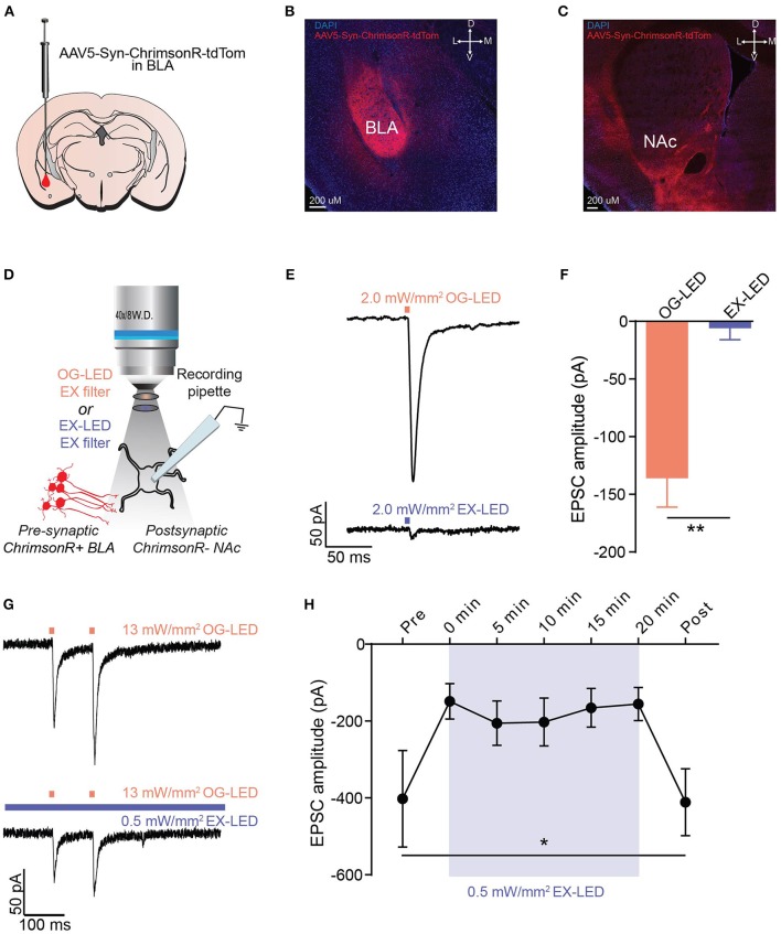
EX-LED has modest effects on pre-synaptic terminals expressing ChrimsonR. (A) Schematic of AAV5-Syn-ChrimsonR-tdTom injection into the BLA. (B) DAPI stained coronal section showing ChrimsonR-tdTom (red) expression in the BLA after virus injection. Scale bar = 200 μm. (C) DAPI stained coronal section showing ChrimsonR-tdTom (red) fiber expression in the NAc after virus injection in the BLA. No evidence of somatic expression of ChrimsonR in the NAc was observed. Scale bar = 200 μm. (D) Optically-evoked EPSCs were recorded from NAc neurons during optical stimulation of BLA-to-NAc fibers with LEDs filtered with the EX- and OG-LED excitation filters. (E) Representative traces showing current in a postsynaptic NAc neuron in response to 2 mW/mm2 of OG-LED stimulation and 2 mW/mm2 of EX-LED stimulation. (F) EPSC amplitude is significantly lower in response to EX-LED stimulation compared to OG-LED stimulation (n = 6 cells from n = 2 mice, paired t-test, p < 0.01). (G) Representative traces showing changes in current in a postsynaptic NAc neuron in response to 13 mW/mm2 pulsed OG-LED stimulation (top) and 13 mW/mm2 OG-LED pulsed stimulation during extended EX-LED stimulation (0.5 mW/mm2). (H) OG-LED light-evoked postsynaptic currents are significantly attenuated during exposure to simultaneous EX-LED (n = 6 cells from n = 5 mice, repeated-measures one-way ANOVA, p = 0.04. None of the Bonferroni multiple pairwise comparisons were significant). Error bar is SEM. *p < 0.05; **p < 0.01.

