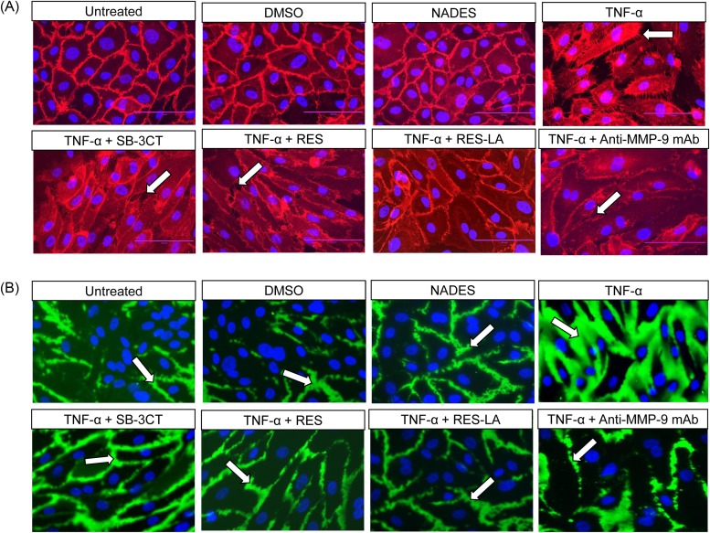
Figure 5. Localization of PECAM-1 and intercellular spaces.
HUVEC (3 × 10⁵ cells/well) were plated for 48 h on biotinylated-gelatin coated chambers. TNF-α (100 ng/ml) was added in the presence or in the absence of 10 µM RES, RES-LA, SB-3CT, or with anti-MMP-9 mAb for 24 h. All samples were treated either with a Cy3-conjugated mouse anti-human CD31/PECAM-1 mAb (A) or with a fluorescein-streptavidin compound (B).

