Figure 2.

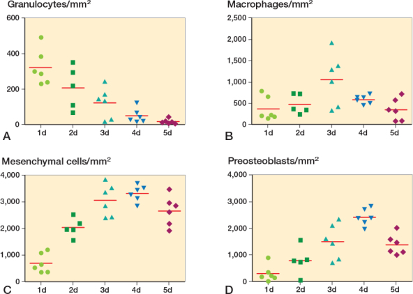
Quantification of cell populations in drill holes in proximal tibia. Granulocytes (A; myeloperoxidase, MPO). Macrophages (B; CD68). Mesenchymal cells (C; vimentin). Preosteoblasts (D; RUNX2).

Quantification of cell populations in drill holes in proximal tibia. Granulocytes (A; myeloperoxidase, MPO). Macrophages (B; CD68). Mesenchymal cells (C; vimentin). Preosteoblasts (D; RUNX2).