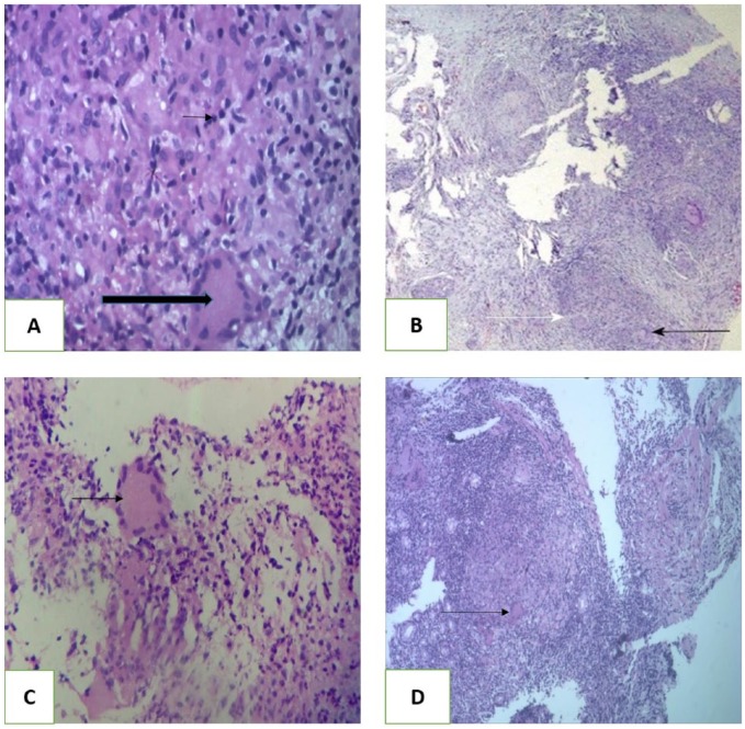
Figure 2.
Histopathologic section (hematoxylin-eosin, original magnification ×100). (A) Case 1—gastric ulcer edge biopsy showing caseating granuloma (large arrow) and Langerhans giant cell (small arrow). (B) Case 2—duodenal erosion biopsy showing granuloma formation (black arrow). White arrow showing Langerhans cell. (C) Case 3—ill-formed Langerhans giant cells with granuloma formation. (D) Case 4—stomach cardia growth biopsy showing caseating granuloma and Langerhans giant cell.

